What Does Glia Do in the Brain?
Glia – Part of the Brain’s Neuroanatomy: What Is It and What Does It Do?
A Three-Part Series: Part I, Section 1
by Rae Marie Gleason
 Learning to live life with a chronic pain illness is not for the faint of heart. It takes personal fortitude and determination along with an inquisitive mind and self-empowerment. And knowledge about the chronic pain illness is the first and foremost element in helping a person regain control of his or her life.
 Changes in brain chemistry go hand in hand with pain felt in the periphery (i.e., leg, arm or lower back). Understanding these changes takes research and learning as much as possible about how the brain perceives and reacts to pain and sickness.
Over the past 20 years scientists have escalated brain chemistry research to better understand what is happening in the brain that results in pain felt in the periphery. This statement may be reversed and expressed as what happens in the brain as a result of an injury in the periphery. It is impossible to separate these two important concepts in the pain process. Intricate scientific studies are looking for understanding of the constant changes and adjustments forced on the brain by a chronic pain condition.
In recent years research has focused on the relevance of glia in the spinal cord and brain, and how it affects the magnitude of a chronic pain condition and a sickness response. This is a vital step in the understanding of chronic pain and its effects on a person’s body. Consider disorders such as fibromyalgia, TMJ, interstitial cystitis, IBS and other pain conditions that are more than pain; they are sickness. Sickness is that awful feeling of just wanting to stay in bed and rest until a person feels better, not wanting nourishment or communication with people, because the effort is just too great. The feeling of nausea can overcome a person who hurts so much that they are physically ill. This is what glia cell research is all about.
Beginning with this July 2014, Advocate Voice Newsletter, the NFMCPA will offer a three-part series introducing glia and its function in the human brain and spinal cord. By learning about glia you can start to gain a better understanding of research in this field and how it can affect you and living better with your chronic pain condition. Knowledge is the key in
- better understanding pain;
- how it affects you physically and emotionally; and
- how to incorporate treatment modalities that are beneficial in helping to relieve your suffering in both these areas.
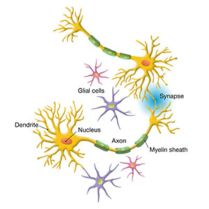
Glial cells are also called neuroglia or glia, which is Greek for "glue." The two main types of cells in the nervous system are glia and neurons. Glial cells are non-neuronal cells which provide support, protection and nutrition for neurons, as well as maintain homeostasis and form myelin. "The major distinction is that glia do not participate directly in synaptic interactions and electrical signaling, although their supportive functions help define synaptic contacts and maintain the signaling abilities of neurons. Glia are more numerous than nerve cells in the brain, outnumbering them by a ratio of perhaps 3 to 1." (Neuroscience, Sinauer Associates, Inc. 2nd Edition, NIH online at http://www.ncbi.nlm.nih.gov/books/NBK10869/)
After glia, we will discuss mitochondria and its relevance to pain and sickness. Three types of glial cells have been discovered in the central nervous system: astrocytes, oligodendrocytes and microglia. Schwann cells, which are similar to glial cells, are abundant in the peripheral nervous system:
- Astrocytes are a heterogeneous cell population, which interacts with neurons and blood vessels.
- Oligodendrocytes in the central nervous system (and Schwann cells in the peripheral nervous system) produce myelin and are responsible for the high speed information processing in the axons of vertebrates.
- Microglial are central nervous system immune cells and react with a supposed activation to changes in the nervous system.
Part I: Introduction and Astrocyte Glia
There are two major types of cells in the brain: neurons and glial cells. While much is known about neurons that have been studied for more than a century, in-depth glial cell research has only developed over the last decade. Over the past few years science has advanced better understanding of brain function and the importance of the interaction of all its cell types, including neurons and glial cells.
For the past two decades glial cell research has considerably increased for several reasons, including studies in molecular biology that have revealed gene products in the brain which are often expressed by these cells. Additionally, animal models have been developed for all major neurological and psychiatric diseases which have helped researchers better understand cellular responses in different pathological stages. Scientific evidence now exists that reveals there is not a single pathological process in the brain that occurs without involvement of glial cells, specifically microglia and astrocytes.
Evidence of glial excitability is key to better understanding cellular brain activity. Neuron responses are known to be quicker; and even though glial cells (and astrocytes in particular) have been shown to communicate more slowly, they have recently been discovered to modulate neuronal activity and ultimately brain function. It is now assumed that the combination of activities of glial cells and neurons is crucial for all brain functions, including thinking, emotions and other functions which define the human spirit.
Glial cells are termed the pathological sensors of the brain. They migrate to the site of damage where they can proliferate and become phagocytes. Glial cells are known to interact with the peripheral immune system by antigen presentation.
In today’s world, science conceives the brain as an organ which can achieve its function due to the interaction of all these cell types only. This is especially true in pathological (persistent) states.
Heterogeneity of Astroglia
The most numerous and diverse neuroglial cells in the central nervous system (CNS), astrocytes are literally “star-like cells.” While neuroscientists are pretty certain they understand what an astrocyte is, there is no uniform and unequivocal definition of these cells:
• Not all astrocytes are star-like cells
• Not all astrocytes express the specific marker glial fibrillary acidic protein (GFAP)
• Not all astrocytes contact brain capillaries
Astrocytes are actually the cell population in the brain which are left over after neurons, oligodendrocytes and microglial cells are removed. Subsequently, astroglial cells display a heterogeneity in their morphology and function. They appear to be as heterogeneous (mixed; different kinds/parts) as neurons. In different brain regions they may have very different physiological properties.
Morphology of Astrocytes
Morphology (branch of biology that deals with the form and structure of animals and plants) is highly differential. Although some astrocytes do have a star-like appearance, many more morphological profiles exist. An archetypal morphological feature of astrocytes is their expression of intermediate filaments, which form the cytoskeleton. Glial fibrillary acidic protein (GFAP) and vimentin are the main intermediate filament proteins in the main types of astrocytes; GFAP is commonly used as a specific marker for the GFAP expression. It works well as a marker in cultured astrocytes, but in situ (encapsulated) the levels of GFAP expression greatly vary. For example, GFAP is expressed by virtually every Bergmann glial cell in the cerebellum but only about 15-20 percent of astrocytes show up in the cortex of mature animal GFAP expression.
Protoplasmic astrocytes are present in the brain’s gray matter. They are comprised of many fine processes (tendrils), which are extremely elaborate and complex. Protoplasmic astrocyte processes contact blood vessels, forming so called “perivascular” endfeet, and form multiple contacts with neurons. Some also send processes to the pial surface where they form “subpial” endfeet. Protoplasmic astrocytes density varies in the cortex and covers most of neuronal membranes within their reach.
Fibrous astrocytes are present in the brain’s white matter. Their processes are long though much less elaborate as compared to protoplasmic astroglia. The processes of fibrous astrocytes establish several perivascular, or subpial endfeet. They send numerous extensions (perinodal processes) that contact axons at nodes of Ranvier.
Adapted from Network Glia, www.networklia.eu/en/microglia
Image courtesy of NIH NICHD https://www.nichd.nih.gov/news/releases/Pages/012811-communication-between-brain-cells.aspx


